Safranin O – the versatile dye for biological and chemical analysis
Safranin O is a cationic dye that plays a central role in biological and chemical analysis. This red dye is used in numerous applications, from staining tissue sections in microscopy to identifying certain organic compounds. In this blog post, we will take a closer look at Safranin O and its diverse applications.
The Chemistry of Safranin O
Safranin O, also known as Safranin T or Safranin 3B, is an organic compound with the chemical formula C₂₀H₁₉ClN₄. It is a cationic dye belonging to the azine dye group. Safranin O has a red color and is highly soluble in water.
The structure of Safranin O consists of an aromatic ring system with a positively charged nitrogen group. This cationic property is crucial for many applications of the dye, as it allows it to bind electrostatically to negatively charged structures.
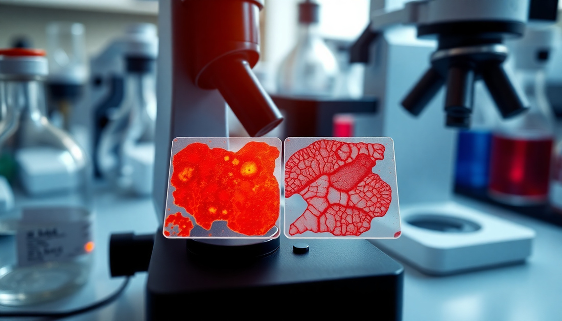
Microscopic staining with Safranin O
One of the main applications of Safranin O is the staining of tissue sections in light microscopy. Safranin O binds selectively to specific structures in cells and tissues, thus enabling high-contrast visualization.
In botany, Safranin O is frequently used to make cell walls and lignifications in plant cells visible. The dye binds to lignin and cellulose and stains these structures red. For example, wood cells in cross-sections of plant tissues can be easily identified.
Safranin O also plays an important role in histology and cytology. Here, the dye is often used in combination with other dyes such as light green or methylene blue to achieve differential staining. For example, cell nuclei, cytoplasm, and extracellular structures in tissue sections can be specifically stained.
Analytical Applications of Safranin O
In addition to microscopic staining, Safranin O also finds diverse applications in chemical analysis. Due to its cationic properties, the dye can bind to various anionic structures and thus be used to detect certain substances.
One example is the detection of sulfates. Safranin O forms an insoluble red complex with sulfate ions, which can be quantified photometrically. This procedure is frequently used in water analysis to determine sulfate levels in water.
The detection of phosphates is also possible with Safranin O. Here, the dye forms a red complex with phosphomolybdic acid, which can also be detected photometrically. This method is used in soil analysis and environmental analysis.
Furthermore, Safranin O can be used to detect proteins. The dye binds to cationic amino acid residues and stains proteins red. This property is used, for example, in electrophoresis to make protein bands visible in gels.
Further Applications of Safranin O
In addition to the described applications in microscopy and analysis, Safranin O is also used in other areas:
Bacterial Staining
Safranin O is frequently used in bacteriology to stain Gram-positive bacteria. Together with crystal violet, it forms part of the Gram staining method, which serves to distinguish between Gram-positive and Gram-negative bacteria.
Textile Dyeing
As an azine dye, Safranin O can also be used for dyeing textiles. The dye binds well to natural fibers like wool and silk and produces a vibrant red color.
Medical Applications
In medicine, Safranin O is used as a dye for diagnostic purposes, for example in urology to visualize urinary tract infections.
Conclusion
Safranin O is a versatile dye that plays a central role in biological and chemical analysis. From staining tissue sections in microscopy to detecting ions in water analysis – Safranin O is indispensable in daily laboratory and research work. Its properties as a cationic dye make it a valuable tool in modern analysis.